UC Berkeley sleep scientists report that as Alzheimer’s-linked tau builds in the frontal cortex, the deep-sleep brain waves that normally sweep across the scalp turn shorter and more solitary — “lonely” waves — and overnight memory consolidation gets worse. The study, led by Omer Sharon with Matthew P. Walker and colleagues, appears in Nature Neuroscience (11 Sep 2026). Participants were cognitively healthy; this is a mechanism bridge between tau and memory via non-REM sleep, not a claim that everyone with lonely waves has Alzheimer’s, and not proof that tau alone causes the memory loss.
Why it matters
Episodic memory — where you parked, what you learned last night — softens with age for almost everyone, and collapses in Alzheimer’s. Researchers already knew tau tangles track cognitive decline. Separately, they knew deep non-REM sleep hosts slow waves: huge populations of neurons switching off and on together as the wave travels across cortex, starting in frontal areas.
This paper stitches those stories. Frontal tau is associated with weaker en masse unity of slow waves and less cortical travel; those sleep features, in turn, track how well people keep word associations overnight. If the bridge holds, sleep physiology becomes a measurable intermediate — potentially cheaper to sample than PET — on the path from molecular pathology to the memory complaint people actually feel.
Key numbers
| Quantity | Value (Nature Neuroscience / Berkeley News, Sep 2026) |
|---|---|
| Peg | Frontal tau ↔ lonely, nontraveling NREM slow waves ↔ overnight memory |
| Paper date | 11 Sep 2026 (Nat Neurosci) |
| Young vs older EEG | early 20s vs mid-60s–mid-70s (cognitively healthy) |
| Young-wave travel (press) | roughly a handspan across the scalp |
| Cortical involvement effect size | N = 42 |
| Slow-wave distance traveled | N = 39 |
| Overnight word consolidation | N = 40 |
| Longitudinal memory change | N = 25 |
| Longitudinal cortical involvement / tau change | N = 19 |
| WashU CSF cohort (MoCA / Craft recall) | N = 82 |
| Longitudinal frontal tau vs cortical involvement | β = –11.78, p = 0.001 (N = 19) |
Primary numbers are from Sharon et al., DOI 10.1038/s41593-026-02415-9, with the handspan comparison from UC Berkeley’s 11 Sep news release. The headline stays on lonely waves and tau; the Ns are what keep it honest.
What they measured
During non-REM sleep, slow waves are not just “deep sleep on a watch.” Each wave is a coordinated shutdown cascading across cortex. Sharon’s group used clinical EEG to quantify how many electrodes join a wave (cortical involvement) and how far the wave travels. In younger adults, clusters moved about a handspan. In older adults, waves traveled less and looked more solitary.
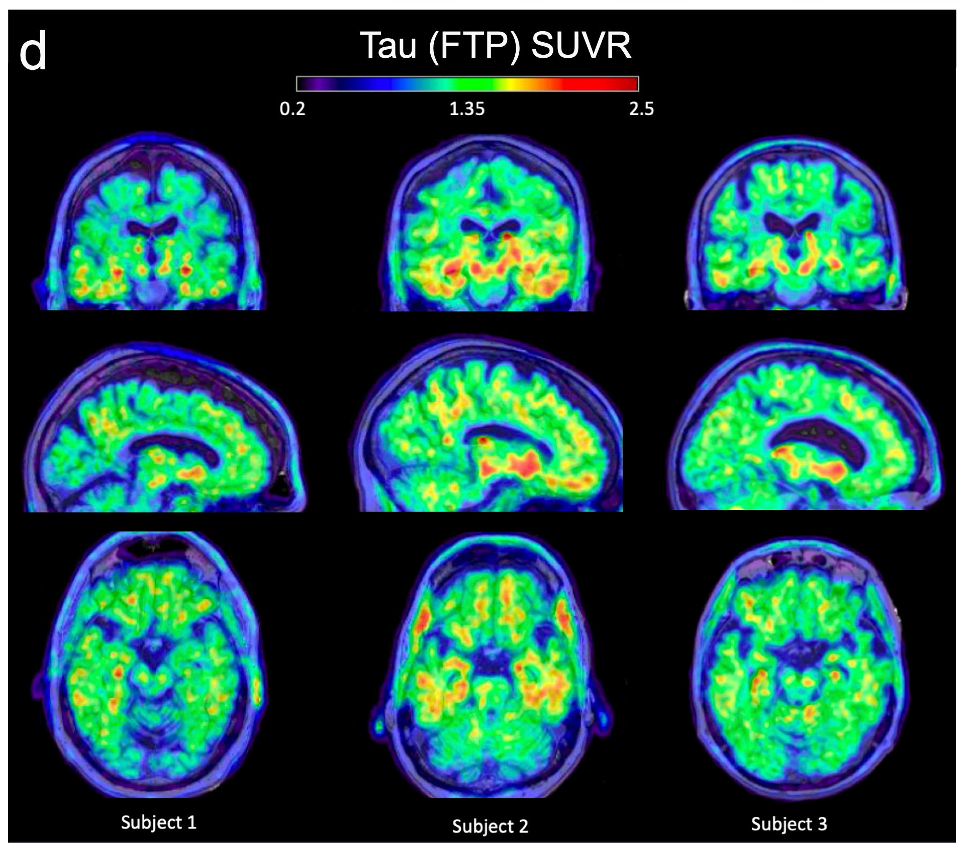
PET with William Jagust’s group mapped tau in the same older participants. More frontal tau lined up with the sleep breakdown. A word-pair task before and after sleep scored overnight consolidation: people whose waves were lonelier and shorter-reaching remembered less the next day.
Years later, a subset returned. Those whose frontal tau burden rose showed steeper drops in cortical involvement and weaker overnight retention. In the N = 19 longitudinal PET window, greater increases in frontal tau associated with reductions in cortical involvement (β = –11.78, p = 0.001). Correlation is not causation — the authors are explicit — but the time course is what you want before you design an intervention.
Because PET is expensive, the team replicated the sleep pattern with cerebrospinal-fluid Alzheimer’s markers in an independent Washington University cohort led clinically by Yo-El Ju (N = 82 on MoCA and Craft recall endpoints in the extended data). Higher CSF tau-related ratios tracked lonelier slow-wave physiology even without frontal-only PET. Two cohorts, two pathology readouts, same sleep story.
What this is not
These volunteers did not have Alzheimer’s disease. Sharon told Berkeley News they had tau with subclinical impacts and memory changes still in the normal aging range. Lonely waves here are an early physiology signal, not a diagnosis.
The study does not prove tau causes memory loss by wrecking slow waves. It shows association, mediation-style links, and longitudinal co-movement. Amyloid PET regional controls in the paper did not light up the same way — useful, still not a full causal graph.
It is also not a consumer sleep-gadget trial. No headband was shown to clear tau. The progress claim is mechanistic clarity: memory decline with frontal pathology may run partly through how deep-sleep waves fail to travel.
What to watch
- Which leads. Sharon’s line — “we can see tau and lonely waves rising together; we cannot yet say which leads” — is the trial-design question. Sleep intervention vs anti-tau vs both.
- Scalable assays. If scalp EEG signatures of lonely waves predict PET or CSF trajectories in larger cohorts, clinics get a cheaper monitoring layer.
- Replication outside Berkeley/WashU. Independent aging cohorts with overnight EEG plus tau PET or plasma/CSF biomarkers need to hit the same β direction.
Phone-screen scoreboard: frontal tau, lonely nontraveling slow waves, and weaker overnight memory — measured in healthy older adults, replicated with CSF, with longitudinal PET N = 19 moving together. That is a clearer map of how Alzheimer’s pathology may steal last night’s memories — not a finished treatment.
Sources
- Sharon, O. et al. Nature Neuroscience (11 Sep 2026): Human tau pathology is associated with lonely, nontraveling slow waves linked to memory impairment — DOI 10.1038/s41593-026-02415-9
- UC Berkeley News, 11 Sep 2026: New UC Berkeley study links deep-sleep loss to Alzheimer’s-related protein buildup (Maya L. Kapoor; handspan / quotes)