On 9 September 2026, a Nature team led from Albert Einstein College of Medicine — with partners at the Allen Institute for Brain Science, Stanford, and others — showed that a rare class of cortical inhibitory neurons can actively push mice into sleep. The cells, called Sst-Chodl neurons, make up less than 1% of cortical GABAergic neurons. When researchers turned them on with a chemogenetic drug (CNO, 0.5 mg/kg) in n = 14 mice, time in slow-wave sleep and REM rose while wake fell (P < 0.001 for each), and sleep arrived sooner. The finding is a mouse-circuit result, not a human sleep pill.
Why it matters
For decades, sleep science has treated the cortex mostly as a passenger: brainstem and hypothalamic switches flip arousal, and the cortex follows with slow waves. That story is incomplete. Growing evidence says cortical circuits also help set sleep pressure and synchrony. What was missing was a sparse, genetically identifiable cell type that both tracks low arousal and causes sleep when driven.
Sst-Chodl cells fill that gap. They co-express somatostatin (Sst) and chondrolectin (Chodl) — and typically Nos1 and the neurokinin-1 receptor — and send long-range axons across large stretches of neocortex. Unlike most local inhibitory neurons, one cell can influence many regions at once. When they fire, the cortex looks more like quiet wakefulness and slow-wave sleep: low-frequency synchrony rises. When experimenters activate them across the neocortex, mice sleep more. That is a stake a stranger can care about: a named cortical switch for sleep, not another vague "brain waves change" paper.
Key numbers
| Quantity | Value (Nature OA PDF / Fig. 5, 9 Sep 2026) |
|---|---|
| Peg | Published online 9 September 2026 (CC BY 4.0) |
| Cell class | Sst-Chodl (Sst + Chodl; also Nos1 / Tacr1) |
| Sparsity | <1% of cortical GABAergic neurons |
| Chemogenetic agonist | CNO 0.5 mg kg⁻¹ (i.p.) |
| Behavior cohort (light phase) | n = 14 mice (11 Sstflp;Nos1creER + 3 Sstflp;Chodlcre) |
| SWS time after CNO | Increased vs vehicle, P < 0.001 |
| REM time after CNO | Increased vs vehicle, P < 0.001 |
| Wake time after CNO | Decreased vs vehicle, P < 0.001 |
| Latency to sleep | Reduced, P = 0.003 |
| SWS bout duration | Unchanged (NS, P = 0.110); more bouts, not longer ones |
| Imaging (superficial layers) | n = 15 mice, n = 97 cells — most active in SWS / quiet wake |
| Lead institutions | Albert Einstein College of Medicine; Allen Institute for Brain Science |
Those figures are taken from the open-access Nature article (DOI 10.1038/s41586-026-10876-y) and its Fig. 5 caption — not invented. The phone-readable stake is a cortical sleep switch; the table is what pays that click.
How they found the switch
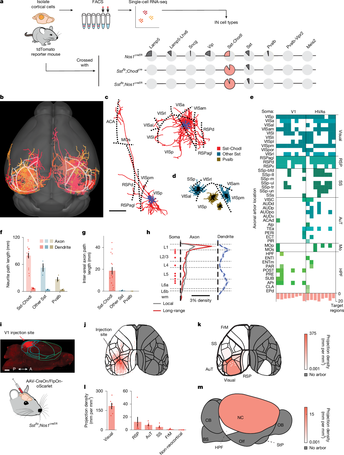
Corresponding authors Geoffrey Terral and Renata Batista-Brito, with co-first author Jacob M. Ratliff, used intersectional genetics so only neurons that hit both Sst and Chodl (or Sst and Nos1) gates got the tools. Morphology reconstructions showed what textbooks rarely emphasize: these inhibitory cells are long-range. Sixteen reconstructed Sst-Chodl cells in visual cortex sent axons far beyond their home area, while comparison Pvalb and non-Chodl Sst cells mostly stayed local. Population tracing from V1 confirmed dense ipsilateral neocortical projections into retrosplenial, auditory, somatosensory and frontal motor territories.
Two-photon calcium imaging then asked when the cells fire. In head-fixed mice, superficial-layer Sst-Chodl neurons were most active during slow-wave sleep and quiet wakefulness and suppressed during movement and REM (ANOVA P < 0.001 across states). Their activity tracked delta-band power — the low-frequency signature of synchronized cortex — and rose just before cortical DOWN states. Deep-layer cells imaged through a microprism told the same story. The cells are not a passive sleep marker; they sit inside the synchrony loop.
Causality came next. Optogenetic activation raised multi-region cortical synchrony. Chemogenetics went further on behavior: an excitatory DREADD (hM3Dq) delivered through 22 neocortical injection sites (11 per hemisphere), then a low systemic CNO dose. In a counterbalanced home-cage design during the light (inactive) phase, CNO increased SWS and REM percentages, cut wake, shortened latency to sleep, and sent mice into the nest with less locomotion — without stretching each SWS bout. Control mice without the DREADD did not show the sleep boost from CNO alone. Dark-phase follow-ups still raised SWS and cut latency, so the effect is not limited to "already sleepy" hours.
What this is not
This is not a human therapy. The entire causal package — intersectional viruses, DREADDs, CNO — is a mouse toolset. CNO pharmacology has its own caveats in sleep studies; the authors cite controls and a dose chosen to limit off-target effects, but translation to people is zero today. It is also not a claim that subcortical sleep centers are obsolete. The paper is explicit: cortical circuits matter alongside established hypothalamic and brainstem mechanisms.
Nor should "<1%" be read as "one cell puts you to bed." The chemogenetic experiment activated Sst-Chodl cells across much of the neocortex. Local stimulation synchronizes nearby networks; the behavioral sleep induction used pan-neocortical drive. And REM rose with SWS even though the cells themselves are quieter in REM — a reminder that network outcomes are not a simple copy of one cell's preferred state.
What to watch
Three near-term checks will show whether Sst-Chodl cells become a standing chapter in sleep neuroscience or a beautiful one-off.
- Independent labs. Can other groups reproduce the P < 0.001 SWS/REM lift and latency cut with the same intersectional lines or Allen enhancer AAVs?
- Human homology. The cell type is described as conserved from salamanders to humans in transcriptomic atlases; watch for invasive or imaging evidence that homologous long-range inhibitory neurons track low arousal in people.
- Disease and drugs. If insomnia, depression or neurodegeneration dysregulate this class, the circuit becomes a target — still a hypothesis, not a pipeline asset.
For a progress scoreboard that fits a phone screen, keep the pair the paper actually shipped: a cortical cell class thinner than 1% of inhibitory neurons, and a chemogenetic flip in 14 mice that raised SWS/REM and cut wake at P < 0.001. Everything else — sleeping pills, closed-loop stimulators, "we solved insomnia" slogans — has to wait for the experiments this circuit now makes possible.
Sources
- Ratliff, J. M., Terral, G., et al. Neocortical long-range inhibition promotes cortical synchrony and sleep. Nature (published online 9 September 2026). DOI 10.1038/s41586-026-10876-y (open access, CC BY 4.0). PDF: nature.com article PDF
- Key stats verified from OA PDF Fig. 5 caption: n = 14 mice; CNO 0.5 mg kg⁻¹; SWS/wake/REM P < 0.001; latency P = 0.003*; sparsity <1% of cortical GABAergic neurons (main text).