Every electron that flies through a transmission electron microscope is a quantum object. Almost every instrument in service still treats it as a classical tick on a detector. A consortium of Austrian universities has published a way to stop throwing the rest of that information away: couple the beam to a small trapped-ion quantum computer sitting on the electron’s path, and accumulate a stronger signal from a weaker dose.
The work is a design plus a proof, not a gallery of sharper micrographs. The paper is in Physical Review Letters (DOI 10.1103/w6t7-9txs) and on arXiv as 2601.11446, submitted 16 January 2026. TU Wien described the project on 18 August 2026. The microscope itself is now being assembled at the University Service Centre for Transmission Electron Microscopy (USTEM) in Vienna.
What happened
On 18 August 2026, TU Wien described a “quantum computer microscope”: an electron beam that interacts with ions held along its path, so that each electron can share a quantum state with a qubit. First author Elias Pescoller, a doctoral student at TU Wien’s Institute for Theoretical Physics and the Institute of Atomic and Subatomic Physics, puts the operating idea in one line: let the electrons interact with those ions, create entanglement, and then use quantum-computing operations to combine information across successive electrons.
The collaborating groups are named, funded, and assigned jobs:
- TU Wien — theory and USTEM, where the instrument will live. Named researchers include Pescoller, Iva Březinová, Philipp Haslinger and Dennis Rätzel.
- Johannes Kepler University Linz — Johannes Kofler’s group, on the algorithms that combine successive electron–ion interactions.
- University of Innsbruck — Philipp Schindler’s trapped-ion processor, which is the quantum computer being integrated into the microscope.
- University of Vienna — Thomas Juffmann; the University of Vienna coordinates the consortium.
Phys.org carried the same account on 23 August 2026. That is the secondary item the live Good Signal article cited — and then failed to open. The Phys.org piece names the labs, the people, the journal, and the preprint. None of that made it into the published post, which instead told readers that “details remain sparse” and that “the report does not name the researchers.”
What the team actually claims, in their own words: conventional atomic-resolution imaging already works, but it needs a lot of electrons, and proteins and other biological samples do not survive that dose. If more information can be taken from each electron, the dose can fall. The ions become a memory for the beam. Quantum processing on that memory is meant to pull a usable signal out of what would otherwise look like noise. Pescoller’s line is that quantum physics lets the method beat the statistical limits of simply counting electrons.
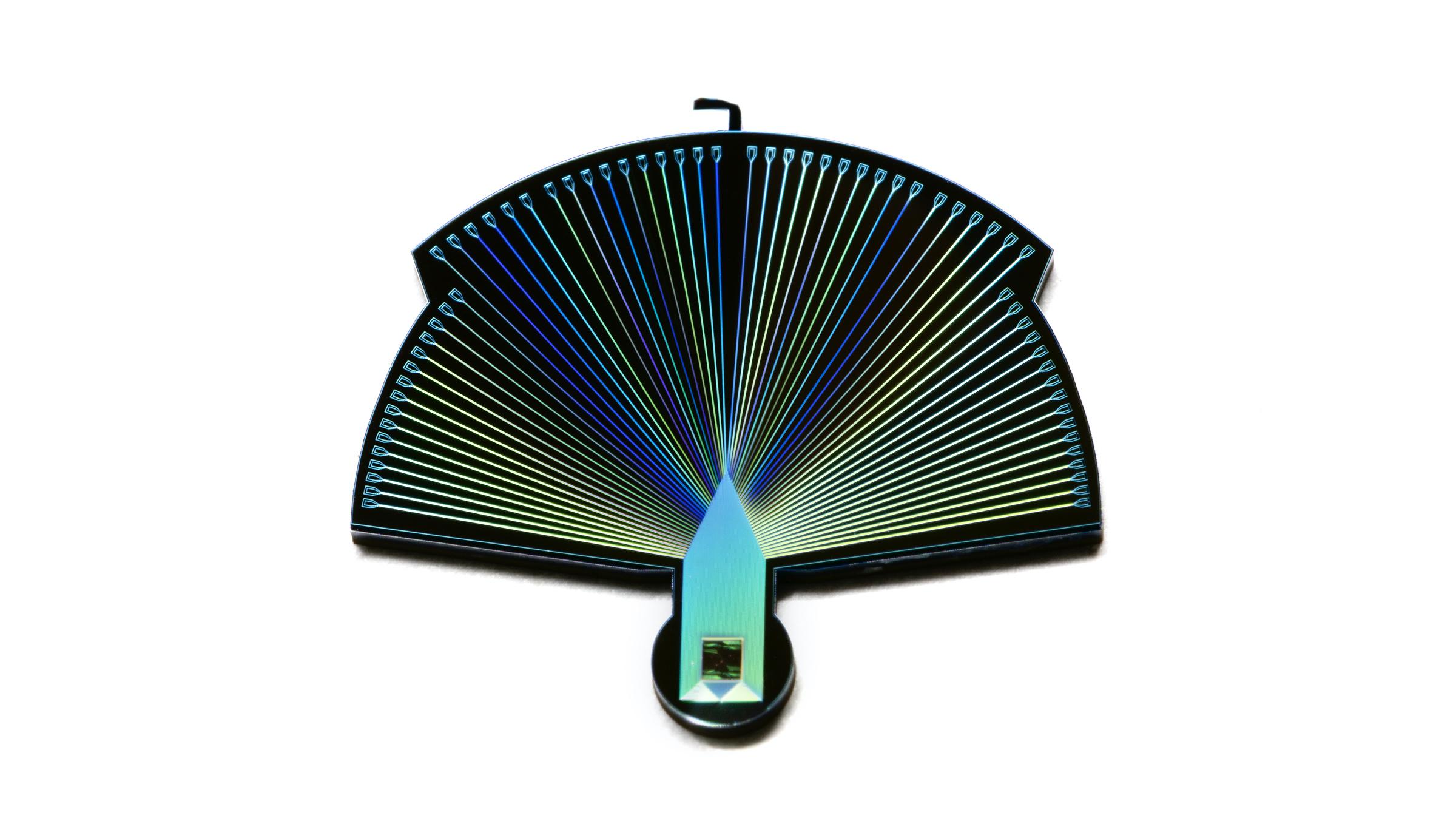
The preprint is specific about the hardware. The ions are calcium-40. They sit in a planar surface-electrode Paul trap, about a hundred micrometres above a photonic chip, with an opening so the electron beam can pass. Typical trap frequencies lie between 0.5 MHz and 5 MHz, corresponding to harmonic-oscillator ground-state sizes of 40 nanometres down to 13 nanometres. The chip that has to fit inside a TEM sample holder is of order 15 mm by 25 mm. The vacuum at the trap needs to reach about 10⁻⁹ mbar or better. Radio-frequency trap potentials can run to 50 volts at around 20 MHz; the authors say the electron pulses must be timed to the zero-crossing of that field so the beam is not deflected.
The coupling is Coulomb, not a new lens. A focused electron imprints a phase on the ion’s motion. If the ion is prepared in a cat state — a superposition of two coherent motional states entangled with the qubit — that phase becomes a rotation on the qubit. After the interaction, the motional states are recombined. For slow electrons (100 eV to 1 keV) and cat-state sizes already demonstrated experimentally (the paper cites |α| = 6.5), the probability of finding the qubit flipped reaches the order of 0.1 to 1. Those energies are not the usual 100 keV TEM setting; the authors point to low-energy electron microscopy, acceleration/deceleration stages, and multi-pass geometries as the routes that make the strongly interacting regime reachable without giving up spatial resolution. Atomic resolution has already been shown down to about 15 keV in other instruments.
Successive electrons multiply. The same ion can interact coherently with many electrons, so phase shifts add. In a multi-path geometry, a diffraction grating could send different electron paths onto different ions, entangling the processor with the path degree of freedom. After the specimen imprints a weak phase, a momentum-space measurement of the electron plus a correction on the qubits transfers that phase onto the register. In an idealised two-path protocol with a full bit-flip coupling, the Fisher information for a specimen phase can scale as n² in the number of electrons — Heisenberg scaling — rather than the classical n. The same appendix is honest about loss: if an electron is lost when the ion and electron are maximally entangled, the accumulated phase is erased. An advantage on average survives only up to a loss probability of about 0.3, and only at a carefully chosen n.
Two limits belong in the lede, not the footer. First, the advantage so far is mathematical. Second, the hardware step is under construction, not commissioned. Haslinger is explicit that today’s microscopes already resolve atomic detail; the bottleneck this project is aiming at is damage, not the existence of atomic resolution.
Funding for the consortium (qcem.info) comes via Austria’s FWF Cluster of Excellence quantA and the Gordon and Betty Moore Foundation (grant GBMF12992).
Why it matters
Electron dose is the quiet constraint on much of structural biology and on a growing list of radiation-sensitive materials. Cryo-EM and related methods already spend extraordinary effort to get more information per electron. A coherent interface between a free-electron probe and a programmable quantum processor is a different attack on the same problem: treat the electrons as quantum sensors, not as a hail of classical particles.
If the USTEM build works, the interesting product is not “a quantum computer that takes pictures.” It is a microscope that can, in principle, run quantum algorithms on the probe itself — entanglement with the ion register, then combining shots — while the sample still sees a conventional electron beam. That is a narrower, more testable claim than the live headline’s “a new way to see the ultra-small.”
It is also a useful counter-example to quantum-computing coverage that only counts logical qubits. Here the processor is small on purpose. Its job is metrology. Innsbruck already runs compact ion processors with more than 15 ions; this project needs that toolbox inside a TEM column, not a larger chip for Shor’s algorithm.
The paper’s other applications — quantum free-electron lasers, nanoscale accelerators, even a generalisation to focused ion beams — are listed as possible extensions. They are not data. The verified object is a TEM–ion-trap interface, on paper, now being built.
What to watch next
- First light of the hybrid column. The next public signal is experimental, not theoretical: an Innsbruck ion trap sitting on the USTEM beam path, and a demonstration that a single electron produces a resolvable qubit excitation — the threshold the preprint argues is reachable at low energy.
- Dose-equivalent images, not slogans. The fair comparison is information extracted per electron (or per unit damage) against a classical TEM on the same sample class, especially proteins. Until those images exist, do not write that the method has beaten the dose limit.
- What does not follow. A PRL on coupling free electrons to trapped ions does not mean atomic-resolution biology is solved, and it does not mean a general-purpose quantum computer has been installed in a microscope. Treat any vendor or aggregator headline that skips those two sentences as a miss.
- The vacuum and timing engineering. The appendices are the watch list: 10⁻⁹ mbar at the trap, electron pulses locked to the RF zero-crossing, a 15 mm × 25 mm chip in a standard holder. Those are the failure modes.
Sources
- TU Wien press release, “The Quantum Computer Microscope,” 18 August 2026 — https://www.tuwien.at/en/tu-wien/news/press-releases/news/das-quantencomputer-mikroskop
- Elias Pescoller et al., “Coupling free electrons to a trapped-ion quantum computer,” Physical Review Letters (2026). DOI: 10.1103/w6t7-9txs
- Same paper on arXiv: https://arxiv.org/abs/2601.11446 (submitted 16 January 2026)
- Phys.org, “Quantum computer microscope is set to significantly improve electron microscopy,” 23 August 2026 — https://phys.org/news/2026-08-quantum-microscope-significantly-electron-microscopy.html
- Consortium site — https://qcem.info