National Eye Institute scientists have published what they call the largest open reference for the size of individual cone photoreceptors — the cells that give you color and sharp central vision — in living human eyes. Nancy Aguilera, Johnny Tam and colleagues report in Investigative Ophthalmology & Visual Science (19 August 2026) a verified set of 9,350 cone inner segments from 28 healthy volunteers, 14 women and 14 men, aged 12 to 84.
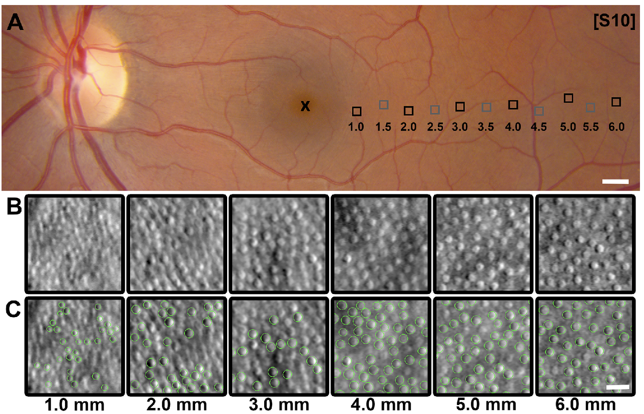
Cone diameter rose with distance from the fovea: 5.0 µm at 1.0 mm eccentricity to 7.8 µm at 6.0 mm. Women’s cones were about 5 percent larger than men’s across most of that strip, a gap the authors say is not explained by axial length or age. Younger people’s cones were generally larger than older people’s. The cells shrink over the decades, most clearly at moderate distances from the fovea.
This is not a treatment. It is a map of “normal,” so that when a clinic images a patient with retinitis pigmentosa or choroideremia, the change can be told apart from ordinary aging.
Why it matters
Diseases that steal daylight vision change the size and shape of cones. Without a public, age-and-sex-stratified reference, a slightly smaller cell on an adaptive-optics image could be pathology, or it could be a 70-year-old’s retina doing what 70-year-old retinas do. Tam, senior investigator in NEI’s intramural program, said there had been no large open dataset for healthy cones across ages, sexes and retinal locations.
The stake is earlier detection and a fairer readout of whether a gene therapy or a cell therapy is actually protecting cones. NEI’s 19 August note is blunt about the next use: track cell-level change as new treatments are tested. Aguilera, first author and an engineer in Tam’s lab, frames the file as open-source infrastructure for that work.
TGS already covers NIH when the lab is named and the number is real. This one is both: intramural NEI, 9,350 cells, a sex effect of about 5 percent.
The print
All figures from the IOVS abstract and the NEI news release, which match.
| Volunteers | 28 healthy (14 female, 14 male) |
| Age | 12–84 years, mean 45.1 ± 20.7 |
| Eyes imaged | one per person |
| Cones measured | 9,350 inner segments |
| Eccentricities | 1.0 to 6.0 mm temporal to the fovea |
| Mean diameter at 1.0 mm | 5.0 µm |
| Mean diameter at 6.0 mm | 7.8 µm |
| Female vs male | about 5% larger in women |
| Age trend | younger generally larger; shrinkage with age, clearest at mid-eccentricity |
Each tracing had to survive review until at least three independent graders agreed. An AI algorithm drew the first outline; people deleted fuzzy cells and added misses.
How they did it
Ordinary cameras cannot resolve a single cone through the living eye. Adaptive optics, borrowed from astronomy, measures the eye’s aberrations and cancels them in real time. Tam’s group used non-confocal split-detection adaptive-optics imaging, which shows the inner segment even when the cell is not waveguiding light the way a textbook cone should. That matters in disease, where reflectivity fails before the cell body is gone.
They imaged a strip of retina from the fovea outward, one eye each. Statisticians then fit a linear mixed-effects model for diameter against sex and age, after accounting for eye length, so a tall myope does not get counted as a “large-cone” phenotype.
The paper is open access in the August 2026 IOVS issue (Vol. 67, Issue 10, article 47). Support: NEI Intramural Research Program. Co-authors include Brian P. Brooks, Wadih M. Zein and Laryssa A. Huryn at NEI, with statisticians at The Emmes Company.
Split-detection imaging is the reason this atlas can exist at all. Confocal adaptive-optics pictures light up cones that still waveguide. In early disease those bright spots wink out while the inner segment is still there. Measuring the inner segment — the metabolically active part of the cell — is how you count a cone that has stopped acting like a fiber optic. The NEI pipeline is built for that: image, auto-trace, triple-grade, then model diameter against eccentricity, age and sex.
Retinitis pigmentosa, Usher syndrome and choroideremia are already known to change cone size and shape. Stargardt disease is on the expansion list. The clinical use is unglamorous and high-leverage: a gene-therapy trial that claims “cones preserved” needs a number for what a 55-year-old woman’s cone should look like at 3 mm from the fovea. Until this file, that number was a lab’s private handful of controls.
What this is not
It is not a 9,350-person study. It is 28 people and a lot of cells. It is not a trial in retinitis pigmentosa, Usher syndrome, choroideremia or Stargardt disease — Tam’s group says those comparisons are next. It does not say women’s vision is better, or that shrinking cones are a diagnosis. Five percent is a population offset, not a clinical cutoff. And 19 August is a week before this desk: the peg is the open atlas landing in the literature this month, not a press embargo this morning.
What to watch
Disease datasets built on the same pipeline. Whether other adaptive-optics labs actually use the file instead of collecting their own 28 controls. Expansion beyond the temporal strip already imaged. And whether a 5 percent sex difference holds in larger samples, or turns out to be this cohort.
Sources
- Aguilera N. et al. In Vivo Human Cone Photoreceptor Inner Segment Diameter Database Using Adaptive Optics Retinal Imaging. Invest. Ophthalmol. Vis. Sci. 67, 47 (August 2026). https://doi.org/10.1167/iovs.67.10.47
- National Eye Institute. NIH researchers build first large-scale database of cone photoreceptor cell size in the living human eye. 19 August 2026. https://www.nei.nih.gov/research-and-training/research-news/nih-researchers-build-first-large-scale-database-cone-photoreceptor-cell-size-living-human-eye